Figure 3.
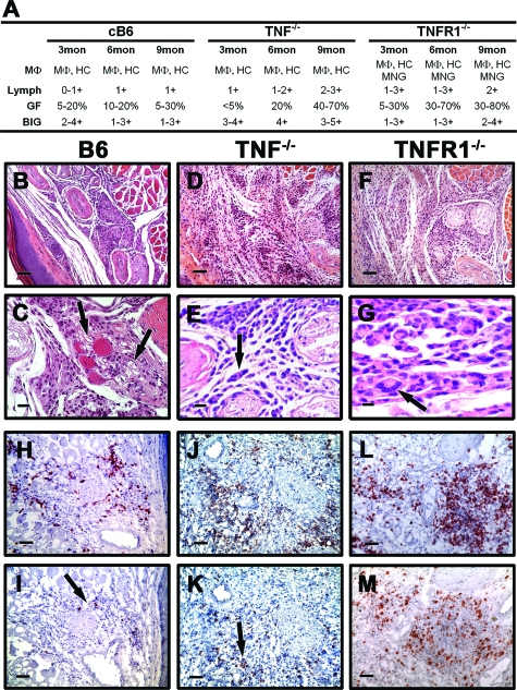
M. leprae-infected TNF−/− and TNFR1−/− FP present a diffuse granulomatous response. B6, TNF−/− and TNFR1−/− mice were infected in each hind FP with 6 × 10³ viable M. leprae. A: Differential granulomatous response was determined by scoring as in Figure 2. B–G: Histopathological (H&E) and (H–M) immunohistochemical staining in FP. B6 FP were comprised of histiocytes, MΦ, and a mild lymphocytic response (B and C, arrows in C); in TNF−/− and TNFR1−/− mice, there was a diffuse and dense infiltration (D and F) with giant cell formation (arrows, E and G). In all three mouse strains, the T cell infiltrate consisted primarily of CD4+ (H, J, and L) cells with a lower influx of CD8+ (arrows, I and K) cells. For (B, D, F, H–M) scale bars = 50 μm; (C) scale bar = 20 μm; and (E and G) scale bars = 10 μm. Experiment shown is representative of two independent experiments.

