Figure 1.
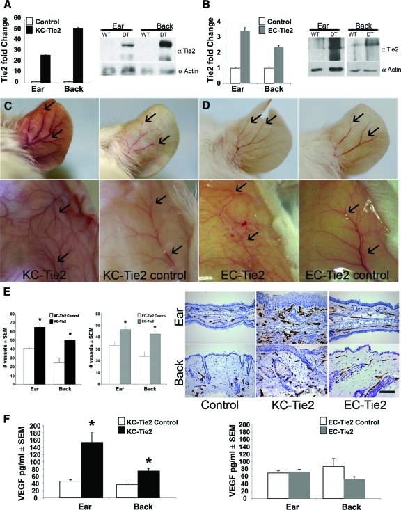
EC-Tie2 and KC-Tie2 mice have distinct phenotypes. Real time RT-PCR and Western blot analysis confirmed increases in Tie2 RNA and protein in ear and back skin of KC-Tie2 (A) and EC-Tie2 (B) double transgenic mice (DT) compared with littermate controls (WT)(n = 6–8 for KC-Tie and littermate controls; n = 4 for EC-Tie2 and littermate controls). KC-Tie2 (C) and EC-Tie2 (D) mice showed increased angiogenesis (arrows) in the ear and superficial fascia compared with control littermates; only KC-Tie2 animals had erythematous ears (C). Endothelial cells of blood vessels in ear and back skin were stained using an antibody specific for MECA-32 (E). KC-Tie2 and EC-Tie2 mice have increased dermal angiogenesis compared with littermate controls (E; n = 4–6 each). KC-Tie2 but not EC-Tie2 mice have increased expression of VEGF protein compared with littermate controls as measured using ELISA (F) (KC-Tie2 n = 10–11; EC-Tie2 n = 7). *P < 0.05 compared with littermate controls. Scale bar = 100 μm.

