Figure 3.
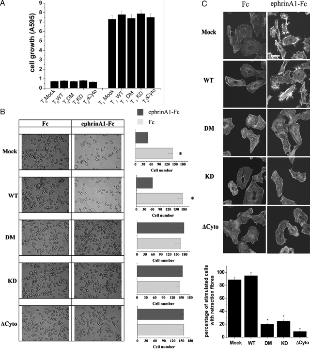
EphA2 phosphorylation is required for ephrinA1-mediated inhibition of ECM adhesion and spreading and for retraction fiber formation. A: Proliferation of PC3 cells transfected with wild-type (WT) and EphA2 mutants. Cells were plated in triplicate in complete medium. Cellular growth was stopped after 4 hours (To) and after 7 days in culture (T7). Cell proliferation was evaluated after crystal violet staining. B: Adhesion assay. PC3 cells expressing wild-type EphA2 or kinase-deficient mutants were kept in suspension for 30 minutes and then seeded onto fibronectin-coated dishes in the presence of 1 μg/ml of ephrinA1-Fc. After 4 hours photographs were taken. The bar graphs represent the number of adhering cells shown in six randomly chosen fields of triplicate experiments. *P < 0.001 stimulated versus control. C: Retraction fiber formation. PC3 cells overexpressing wild-type EphA2 or kinase-deficient mutants were seeded onto collagen-coated coverslips and adhesion was allowed for 24 hours. Cells were serum-starved for 24 hours before stimulating with 1 μg/ml of ephrinA1-Fc for 15 minutes. Confocal microscope analysis after phalloidin-tetramethyl-rhodamine isothiocyanate immunostaining was shown. The results are representative of at least three experiments. The bar graph represents the percentage of ephA1-stimulated cells with retraction fibers, in six randomly chosen fields of triplicate experiments. *P < 0.001 stimulated clones versus stimulated mock. Scale bar = 20 μm.

