Figure 2.
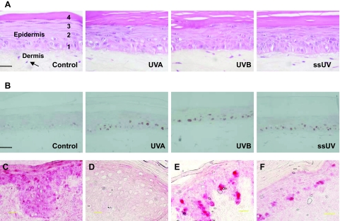
UV radiation does not induce detectable morphological changes in EHS (A). Fibroblasts (arrow) encased in a type I collagen lattice provides dermal support for the epidermis. EHS collected on day 11, 3 days after the final of four UV exposures, were fixed and H&E-stained. Distinct basal (1), spinous (2), granular (3), and cornified (4) layers are recognizable in un-irradiated control, 12.5 J/cm2 UVA-, 0.1 J/cm2 UVB-, and 1.4 J/cm2 ssUV- irradiated EHS. Sections from the same groups of EHS were immunostained for p53 protein (B). Anti-CPD immunostaining in adult human skin collected 24 hours after UV irradiation (C) and in UV-irradiated EHS used for mutation detection (D). Anti-Ki-67 immunostaining in UV-irradiated adult human skin (E) and in UV-irradiated EHS (F). p53 positive cells are stained brown. CPD and Ki-67 positive cells are stained red. Scale bar = 25 μm.

