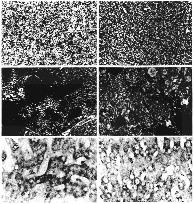
Fig. 7.
Changes in a liver three days after a splanchnic division experiment of group 2 as shown by light microscopy, electron microscopy and autoradiography. The hepatocytes in the nutrient enriched lobes, panels on right, are atrophic; depleted of glycogen and rough endoplasmic reticulum and contain increased smooth endoplasmic reticulum when compared with the enlarged and ultrastructurally normal liver cells in the hormone influenced lobes, panels on left. The rate of cell division, as indicated by autoradiography, is increased on both sides of the liver, but particularly in the hormone influenced lobes. Upper panels, hematoxylin and eosin, ×120; middle panels, electron micrography, ×1,700; and lower panels, autoradiography, ×300.

