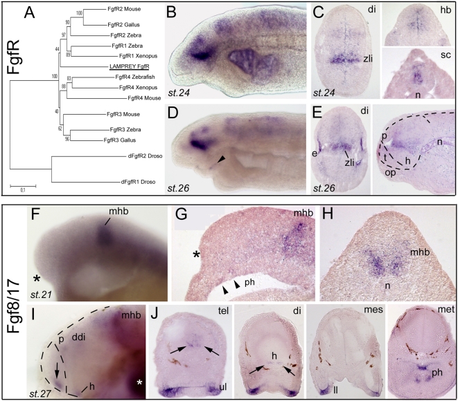
Figure 6. Developmental expression of Fgf signaling components in lamprey forebrain.
A, phylogenetic tree (NJ) of FgfR clones. A contig was assembled out of 7 independent clones (see Table 1), and analysis clearly shows that the unique FgfR present in our database emerges at the base of the tree, and cannot be assigned a robust orthology. B,D (toto) and C,E (sections) show expression of lamprey FgfR. Toto pictures are taken from clones 15 and 83, while section pictures are taken from clones 15 and 16. The right panel in E shows a saggital section, with dotted lines delineating the brain and telencephalon. F,I (toto) and G,H,J (sections; G saggital, H and J coronal) show Fgf8/17 expression (cDNA gift from Kate Hammond [46]). The asterisks in F and G point to the absence of Fgf at rostral telencephalic level at st. 21, whereas the mhb expresses strongly the transcript. Also note faint expression in the presumptive pharynx (arrowheads). The arrows in I and J points to rostral telencephalic expression at the rostral midline at stage 27. At st. 27, strong labeling is also present in the lips and pharynx. White asterisk in I: background trapping in branchial arch. ddi, dorsal diencephalon; h, hypothalamus; p, pineal gland; ph, pharynx.

