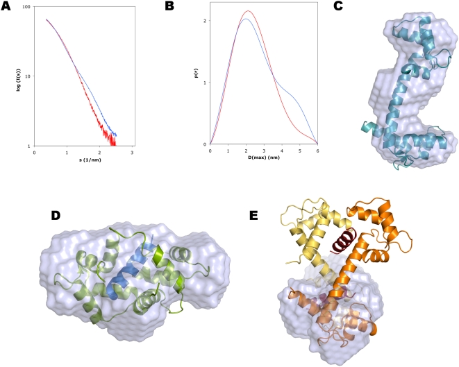
Figure 3. Small-angle X-ray scattering analysis of the solution properties of the complex.
A. Scattering curve. Superposed are the curves for CaM (blue) and the complex (red). B. Distance distribution function. Colouring as in A. Note the disappearance of the shoulder around 4.5 nm in the presence of the peptide. The shoulder is characteristic of the dumbbell-shaped conformation of unliganded CaM, while the distance distribution for the complex indicates a more globular shape. C. Ab initio model of CaM, with the crystal structure of unliganded CaM superimposed. D. Superposition of the ab initio complex solution structure with the crystal structure of a canonical CaM-peptide complex (PDB code 1WRZ). E. Superposition of the SAXS structure with half of the crystal structure of the complex. Colouring as in Figure 1A.

