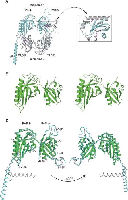
Figure 2. Crystal Structures of Drosophila PERIOD.
(A) Ribbon presentation of the dPER[232–599] dimer with molecule 1 shown in cyan, molecule 2 in grey. The inset shows a close-up view highlighting residues Trp482 and Met560, which have been mutated in this study. The per L mutation site (Val243) is also highlighted.
(B) Stereo view of the dPERΔαF[232–538] monomer structure.
(C) Superposition of the dPERΔαF[232–538] monomer structure (green) on molecule 1 (cyan) and 2 (grey) of the dPER[232–599] dimer. The two orientations are related by 180 ° rotations.

