
3.
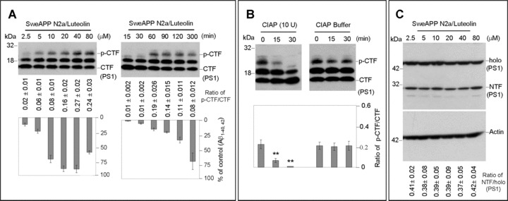
PS1 phosphorylation is associated with luteolin-mediated inhibition of Aβ generation. SweAPP N2a cells were treated with luteolin at a range of concentrations for 4 hrs or at 20 μM for various time-points as indicated. Cell lysates were prepared from these cells and subjected to Western blot analyses of PS1 C-terminal fragments (CTF) (A) and N-terminal fragment (NTF) (C). Western blot analysis by anti-PS1 CTF antibody shows two bands corresponding to phosphorylated PS1 CTF (p-CTF) and one dephosphorylated PS1 CTF (CTF). While Western blot analysis by anti-PS1 CTF antibody shows tow bands corresponding to holo PS1 and PS1 NTF. For (B), cell lysates from luteolin treated cells (20 μM) were incubated with calf-intestine alkaline phosphatase (CIAP) or buffer for various time-points. Western blot analysis by anti-PS1 CTF antibody confirms two higher molecular weight bands corresponding to phosphorylated isoforms. Densitometry analysis shows the ratio of PS1 p-CTF to CTF below figures. A t-test revealed a significant deference between luteolin concentrations and time-points for ratio of PS1 p-CTF to CTF (P < 0.005 with n= 3 for each condition, but not for ratio of holo PS1 to PS1 NTF (P > 0.05 with n= 3 for each condition) at each time-point examined. Cultured media were collected for Aβ ELISA. Data correspond to percentage of Aβ1–40, 42 peptides secreted 4 hrs after luteolin treatment relative to control (untreated) as indicated below panel (A).