
4.
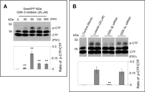
GSK-3α regulates PS1 phosphorylation. For (A), SweAPP N2a cells were treated with a known GSK-3 inhibitor (SB-415286) at 20 μM for various time-points. Western blot analysis by anti-PS1 CTF antibody produces a similar phosphorylation profile to that of luteolin-treated cells. Densitometry analysis shows the ratio of PS1 p-CTF to CTF and ratio of holo PS1 to actin as indicated below the figures. A t-test revealed significant differences between time-points for the ratio of PS1 p-CTF to CTF (P < 0.001 with n= 3 for each condition). For (B), expression of PS1 C-terminal fragments was analysed by Western blot in cell lysates from SweAPP N2a cells transfected with siRNA targeting GSK-3α, β, or mock transfected 48 hrs after transfecion. Prior to experiments, siRNA knockdown efficiency >70% for GSK-3α, β was confirmed by Western blot analysis (data not shown). Densitometric analysis reveals the ratio of PS1 p-CTF to CTF as indicated below the each panel. A t-test revealed significant differences between GSK-3α siRNA-trans-fected cells and GSK-3β siRNA or control (Mock transfected cells) (P < 0.001 with n= 4 for each condition) on the ratio of PS1 p-CTF to CTF. In addition, a t-test also revealed significant differences between luteolin-treated cells and GSK-3β siRNA or control (Mock transfeced cells) (P < 0.001 with n= 4 for each condition) on the ratio of PS1 p-CTF to CTF.