
6.
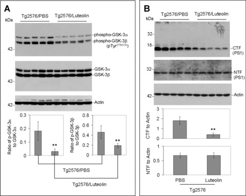
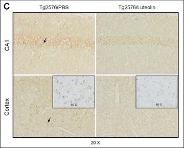
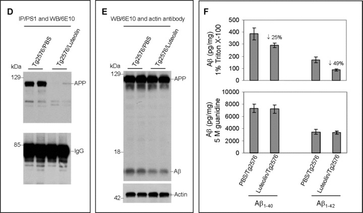
Luteolin reduces GSK-3 activation and cerebral amyloidosis in Tg2576 mice. Brain homogenates and sections from Tg2576 mice treated with luteolin (n= 5) or vehicle (PBS, n = 5). For (A), homogenates were analysed by Western blot with active and holo anti-GSK-3 antibodies with anti-actin antibodies as an internal control. Densitometric analysis reveals the ratio of active phosphorylated GSK-3α/β to holo GSK-3. A t-test reveals significant reductions in both active GSK-3α and β isoforms from luteolin-treated animals compared to control (P < 0.001). For (B), homogenates were analysed by Western blot with anti-PS1 CTF or NTF antibody. Densitometric analysis produces the ratio of PS1 CTF or NTF to actin (internal control). A t-test shows significant reductions in PS1 CTF levels with luteolin treatment (P <0.001), but not for PS1 NTF levels (P >0.05). For (C), immunochemistry staining analysis for active phosphorylated GSK-3α/β. For (D), homogenates were immunoprecipitated by anti-PS1 CTF antibody. Densitometric analysis of Western blot by 6E10 antibody shows the ratio of APP to IgG. A t-test revealed significant differences between luteolin treatment and control (P < 0.001). For (E), homogenates were analysed by Western blot by 6E10 antibody. Approximately 12-kD band may represent oligomeric form of amyloid. Densitometric analysis of Western blot by anti-actin antibody reveals no significant changes in the ratio of APP to actin. For (F), soluble and insoluble Aβ1–40, 42 peptides from homogenates were analysed by ELISA. For Aβ ELISA, data are represented as picograms of peptide present in milligrams of total protein. Luteolin treatment results in markedly reduced soluble Aβ1–40, 42 levels, 25% and 49%, respectively (top panel). No significant reductions in insoluble Aβ isoforms following treatment were observed (bottom panel).