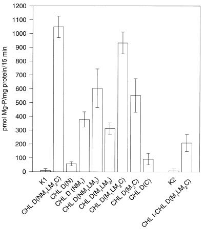
Figure 4.
Diagram of the Mg-chelatase activity of the CHL D constructs (see Table 1) in the presence of CHL I and CHL H. The activity is given in pmol Mg-protoporphyrin IX (Mg-P)/mg protein per 15 min. The SDs were calculated from three independent experiments. K1 and K2 indicate the controls showing the Mg-chelatase activity from yeast cells expressing CHL I and CHL H and solely expressing the artificial I-D fusion protein, respectively.

