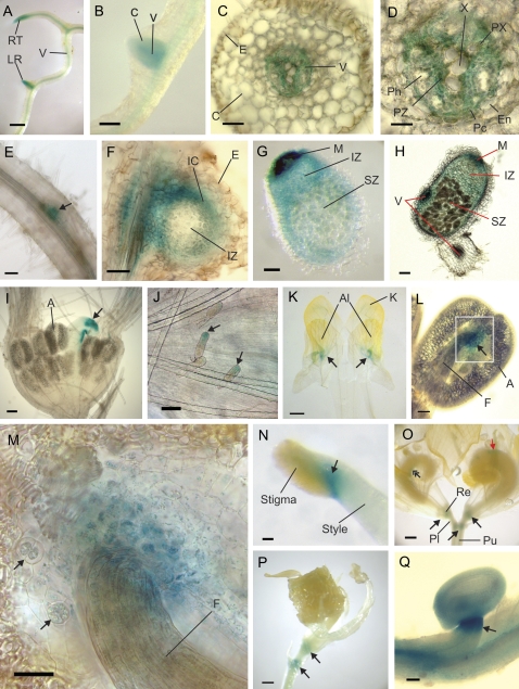
Fig. 2.
prSERK-driven GUS expression during root, nodule, and flower development. (A) In the root there is generalized expression in the vascular tissue with up-regulated expression at the root tip and the site of lateral root formation. (B) Emerging lateral roots show expression in both the vascular tissue and the cortex. (C) Cross-section of a mature root shows that the cortical expression of the emerging lateral root has been lost. Expression is limited to the vascular tissue. (D) A closer view of the root vascular tissue shows expression in the pericycle and the procambial tissue. (E) A hairy root from A. rhizogenes transformation shows lower expression than in that observed in A. tumefaciens-transformed regenerated plants and their progeny. Expression is observed at the site of lateral root formation (arrow). (F) Section through a root nodule, 3 weeks after inoculation with rhizobia showing expression around the vascular tissue and the inner cortex but not in the epidermal cells or infection zone. (G, H) In the mature nodule, expression can be seen throughout the nodule, except in the epidermal cells, but is strongest in the meristem and vascular tissue. (I) The floral meristem shows no expression in the male tissues, with the only expression coming from the developing pistil (arrow) and the glandular trichomes (arrows) on the external part of the meristem (J). (K) Flattened petals from a bud with an incision separating the two halves of the keel petal. Expression occurs in the region where the alae petals (on top) join the keel (arrows). (L) In the stamen there is expression at the junction point where the anther joins the filament (arrow). (M) A closer view of the boxed region in (L), showing expression at the anther/filament junction. Two cells can be seen to be in division (arrows). (N) Expression at the junction of the stigma with the style (arrow) in the female reproductive organs. (O) Expression in the flower occurs at major junction sites of the peduncle with the pedicel and the pedicel with the receptacle (arrows). There is also expression at the adaxial suture line of the ovary (double arrow) and sometimes on other parts of the ovary wall (red arrow). (P) Expression at the major junction sites of the flower persists as the seedpod develops (arrows). (Q) The older flower shows expression in the ovary wall and ovules, with up-regulated expression at the junction site between the ovule and ovary wall (arrow). Scale bars: (A, K, O, P) bar=0.5 mm; (B) bar=0.2 mm; (E, G, H, I) bar=100 μm; (C, F, J, L, N, Q) bar=50 μm. (D, M) bar=25 μm. A, anther; Al, alae petal; C, cortex; E, epidermis; En, endodermis; F, filament; IC, inner cortex; IZ, infection zone; K, keel petal; LR, lateral root; M, meristem; Pc, pericycle; Ph, phloem; Pl, pedicel; Pu, peduncle; PX, protoxylem; PZ, procambial zone; Re, receptacle; RT, root tip; SZ, symbiotic zone; V, vascular tissue, X, xylem.

