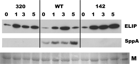
Fig. 8.
Representative Western blot analysis of ELIP and SPPA protein in sppA-320, WT, and sppA-142 leaves. Detached leaves were floated on water in an ice-bath in full sunlight for 4 h, then were allowed to recover in LL at 21–22 °C for the indicated number of hours (0, 1, 3, 5). Membrane proteins were isolated from leaves at the indicated time points for the Western blots (N=3 leaves per time point per genotype). Anti-ELIP1 antiserum was used to probe ELIP levels (top panel) and anti-SPPA antiserum was used to measure SPPA levels on the same blot (middle panel). The gel was loaded on an equal fresh weight basis. A portion of the Ponceau S-stained blot was imaged (M; bottom panel) to show protein loading and transfer (rightmost lane is ∼50 kDa band of the BenchMark Protein Ladder).

