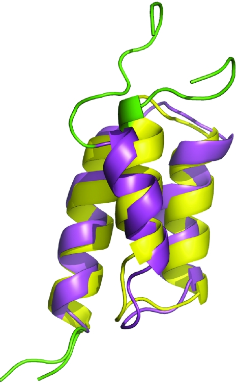
Figure 1.
Structure comparison of the natural B domain (magenta, PDB code 1BDC, first model) and its A2V∕G30A double mutant (Z domain, yellow, PDB code 1Q2N, first model) of protein A. The unstructured regions (residues 1–9 and 57–60) are shown in green color. The overall Cα RMSD between these two structures is 1.7 Å for residues 10–56 and the major difference is the tilt angle of the first helix.

