Figure 1.
sll1 Has Extremely Incurved Leaves, with Deficiency of Sclerenchymatous Cells at the Abaxial Side.
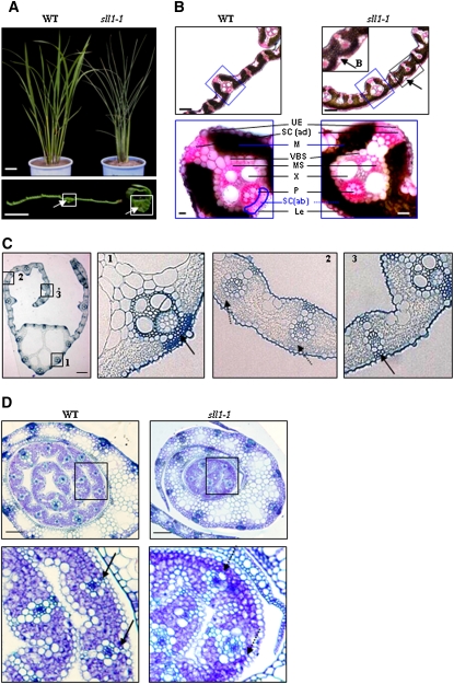
(A) Morphology of wild-type and sll1-1 plants and leaves. Mature wild-type and sll1-1 mutant plants were observed. The clear cells of the midrib (white box) are highlighted with arrows. Bars = 5 cm (top panel) or 2 mm (bottom panel).
(B) sll1-1 leaf blades display altered cellular organization. Bulliform cells on the abaxial surfaces of sll1-1 leaves are highlighted (top panel, arrows). The altered differentiation and distribution of mesophyll cells are highlighted (blue box) in the bottom panel. LE, lower epidermis; SC (ab), abaxial sclerenchyma; B, bulliform cells; P, phloem; X, xylem; MS, mestome sheath; VBS, vascular bundle sheath; M, mesophyll cells; SC (ad), adaxial sclerenchyma; UE, upper epidermis. Bars = 100 μm (top panel) or 20 μm (bottom panel). The broken line indicates the defective sclerenchyma at the abaxial epidermis. Sections (∼20 μm) of rice leaf blade were stained in Ruthenium red solution.
(C) The defective sclerenchymatous cells on the abaxial side of sll1-1 leaves. The defect is mainly in the small veins where the curl occurs (2; broken lines). Cellular organization at the midrib region (1) and the margin of the blade (3) in sll1-1 is normal. Bar = 50 μm.
(D) Transverse section of young seedlings at 5 d after germination. Differentiated sclerenchymatous cells on the wild-type abaxial epidermis are highlighted (left panel, arrows) but still lack a thickened secondary wall. These are deficient in sll1-1 (broken arrows). Bars = 100 μm.

