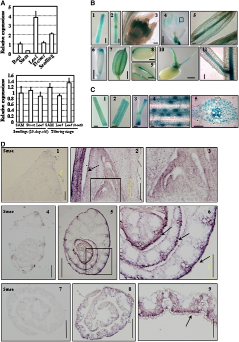
Figure 5.
SLL1 Expression Pattern Analysis.
(A) qRT-PCR analysis of SLL1 expression in various tissues. Expression of SLL1 in roots (from 10-d-old seedlings), stems, leaves (middle part of 4th leaf), flowers, and 10-d-old seedlings (whole plants including roots and shoots) (top panel) in the SAM, shoot (whole plant without roots), and the 4th leaf of 10-d-old seedlings and in the SAM, 10th leaf, and leaf sheath of plants at the tillering stage (bottom panel) are analyzed. For SAM samples, the precisely defined intact SAM and young leaf primordia (without leaf sheath) were collected. Experiments are biological replicates.
(B) Promoter-reporter gene (GUS) fusion studies on the expression of SLL1. Expression of SLL1 in stem (1 and 2), anthers (3 to 5), pistil tip (6), seed vascular tissues (7 to 9), coleoptile and embryonic roots of germinating seeds (10), and root vascular tissues (11) is analyzed. Bars = 1 mm (4 and 6), 2 mm (1, 2, and 7 to 10), or 100 μm (3, 5, and 11).
(C) Promoter-GUS fusion studies of expression of SLL1 in leaf. Expression of SLL1 in young leaf (middle part of 2nd leaf of 10-d-old seedling; 1), mature leaf and leaf vein (2), leaf sheath (3), guard cells (4), and vascular tissues (5) is analyzed. Bars = 1 mm (1), 5 mm (2 and 3), or 50 μm (4 and 5).
(D) In situ hybridization analysis of expression of SLL1 in the abaxial cell layer and in tracheal elements throughout leaf development. The SLL1 signal was detected at the shoot apical meristem and leaf primordia (2; enlarged in 3, arrows). Cross section of the shoot apex showed higher expression of SLL1 in the abaxial cell layer, including the epidermis and vascular system of early leaf blades (5; enlarged in 6, arrows). In the mature leaf, there is an accumulation of SLL1 in the region of the developing abaxial sclerenchymatous cells and tracheal elements (8 and 9, arrow). Arrows highlight the more intense accumulation of SLL1 at the abaxial cell layer. The sense probe was hybridized and used as control (1, 4, and 7). Bars = 200 μm (1 to 4 and 6 to 8), 505 μm (5), or 100 μm (9).

