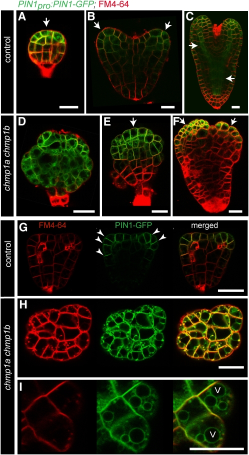
Figure 6.
PIN1-GFP Expression and Localization in Control and chmp1a chmp1b Mutant Embryos. (Embryos were stained with FM4-64 [red] to visualize the cell outlines.)
(A) to (C) Control embryos. Note PIN1-GFP expression at apical/central region (arrow) of a globular stage embryo (A), tips of developing cotyledon (arrows) in heart stage embryo (B), and procambial strands (arrows) of torpedo stage embryo (C).
(D) to (F) chmp1a chmp1b mutant embryos with altered PIN1-GFP expression pattern. Arrows indicate the areas with higher PIN1-GFP expression.
(G) Control heart stage embryo. PIN1-GFP localizes to the plasma membrane in the emerging cotyledons, predominantly to the apical side of the cells (toward the tip of the cotyledons; arrowheads).
(H) and (I) chmp1a chmp1b mutant embryo dissected from the same silique used in (G). Note the substantial PIN1-GFP signal from vacuolar membranes and FM4-64–stained compartments. V, vacuole.
Bars = 20 μm.

