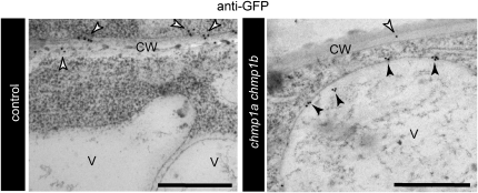
Figure 7.
Immunogold Localization of PIN1-GFP.
Immunogold labeling was performed on high-pressure frozen/freeze-substituted wild type–looking (control) and chmp1a chmp1b mutant embryos from self-pollinated CHMP1A/chmp1a chmp1b/chmp1b/ PIN1pro:PIN1-GFP/PIN1pro:PIN1-GFP plants using polyclonal anti-GFP antibodies. Bars = 500 nm.
(A) Control embryo. GFP signal is detected at the plasma membrane (white arrowheads).
(B) chmp1a chmp1b mutant embryos. White arrowheads indicate gold labeling on the plasma membrane and black arrowheads on the vacuolar membrane. CW, cell wall; V, vacuole.

