Figure 8.
Immunogold Detection of PIN1-GFP and the Endosomal Marker RHA1/RabF2A in Control and Mutant MVBs.
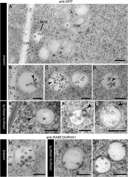
(A) to (G) Immunolabeling of GFP in high-pressure frozen/freeze-substituted WT-looking (control) and chmp1a chmp1b mutant embryos expressing PIN1-GFP. CW, cell wall; TGN, trans Golgi network.
(A) Overview of a control embryo cell showing PIN1-GFP signal on the trans Golgi network, MVBs, and plasma membrane (white arrowheads).
(B) to (D) Detail of control MVBs with gold labeling on MVB lumenal vesicles (black arrowheads).
(E) to (G) Detail of chmp1a chmp1b mutant MVBs. Most of the gold labeling is on the MVB limiting membrane (black arrowheads) and not on MVB lumenal vesicles.
(H) to (J) Immunolabeling of RHA1/RABF2A on control (H) and chmp1a chmp1b mutant MVBs ([I] and [J]).
Bars = 200 nm.

