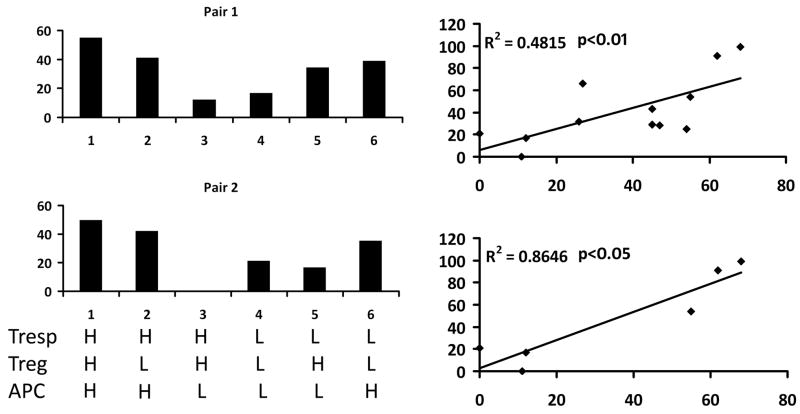
Fig. 5.
Dissection of cellular components critical for in vitro suppression activity. (a) Treg, APC and responder T cells (Tresp) are purified from pairs of subjects, one with high suppression activity (H) in autologous suppression assay and another with low (L) autologous suppression activity. All six combinations of the three cellular components are tested for in vitro suppression activity. These results indicate APC as the possible cell population related to suppression activity. (b) Treg and Teff cells were purified from one blood donor with high autologous suppression activity (T cell donor) and APC were purified from 12 different blood donors with different autologous suppression activity. The function of APC was tested by heterologous suppression assays that mix Treg and Teff cells from the common T cell donor and APC from each of the 12 APC donors. APC function (heterolgous suppression) is plotted against autologous suppression activity.

