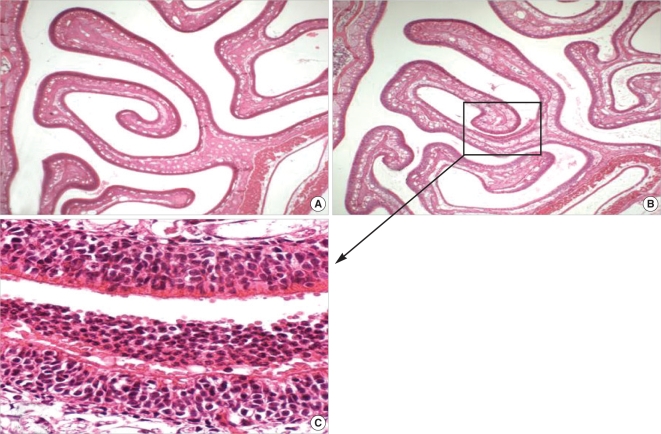
Fig. 1.
Light micrographs of rats sacrificed at day 1. (A) Sinonasal air space bounded by septum, upper nasoturbinate and lower maxilloturbinate in section of control rats shows no evidence of inflammation (H&E, original ×40). (B) Inflammatory cell clusters are observed in sinonasal space of SEB-applied rats (H&E, original ×40). (C) Magnification of marked area on B reveals that the inflammatory cells in sinonasal air space are neutrophils (H&E, original ×400).

