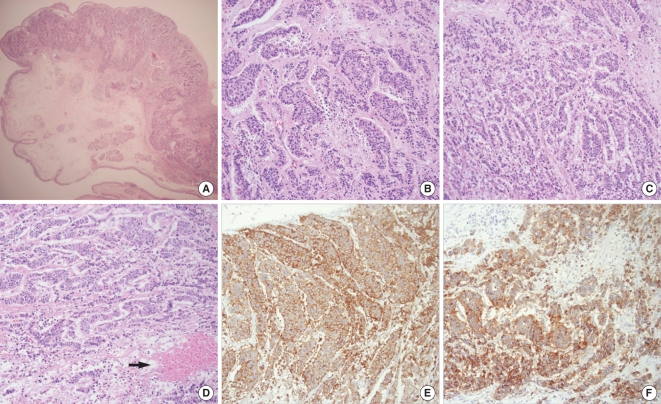
Fig. 2.
Pathologic pictures. (A) Low power view with overlying intact mucosa (hematoxylin-eosin [H&E], magnification ×20). (B) High-power view showing pseudogland and rodget arrangement as a neuroendocrine differentiation (H&E, magnification ×200). (C) Cord and trabecular arrangement with nuclear and cellular pleomorphism (H&E, magnification ×200). (D) Tumor necrosis area (arrow) (H&E, magnification ×200). (E) Positive immunoreaction to chromogranin (magnification ×200). (F) Positive immunoreaction to synaptophysin (magnification ×200).

