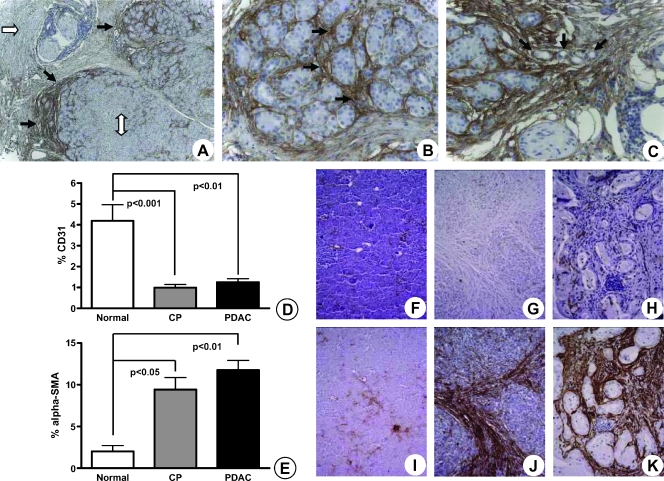
Figure 2.
Site-specific deposition of periostin-rich stroma on the invasive front of the activated stroma in PDAC parallels increased α-SMA expression of PSCs and decreased vascularity in the diseased pancreas. Compared with both normal parenchyma (A; original magnification, x50; white double-headed arrow) and organized ECM (white arrow), periostin expression dramatically increased at the interface where the activated stroma bordered on normal acini. The activation of stromal cells and detection of periostin expression in the CP-like changes surrounding the cancer occur even in areas where no cancer cells are visible. Although some periostin staining was detected in the interlobar septa (B and C; original magnification, x200), the strongest expression was detected in the periacinar spaces (B, arrows). Notice the encasement of acini by the periostin-rich ECMand emergence of tubular complexes (C, arrows; original magnification, x200). The percentage of CD31 (D) and α-SMA (E) staining in normal pancreas, CP, and PDAC tissues is graphically depicted. Results are expressed as mean ± SEM. Evaluation of CD31 (F, G, H) and α-SMA (I, J, K) using consecutive sections of normal (F, I), CP (G, J), and PDAC (H, K) tissues is shown.

