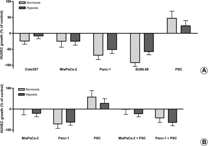
Figure 5.
Assessment of HUVEC growth after treatment with PCC and PSC SNs: HUVECs were seeded in 96-well plates (5000 cells per well) in complete endothelial cell growth medium (100 µl per well). Twenty-four hours later, 100 µl of cancer cell or stellate cell or coculture SN was added to the cells. Forty-eight hours later, cell growth was assessed by MTT assay corrected for day 0. The experiments were performed at least nine times using three different HUVEC clones and three different PSC clones. The effect of four different cancer cell lines and PSC SNs on HUVEC growth is shown in panel (A). The effect of the common SN after coculture of MiaPaCa-2 and Panc-1 with PSC is shown in panel (B). The growth-inhibiting or growth-promoting effects of SNs on HUVECs are shown as percent change of the control (0%).

