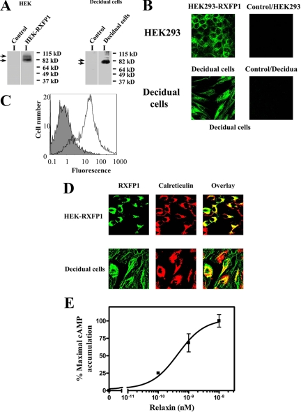
Figure 1.
A, RXFP1 protein expression in HEK-RXFP1 and primary decidual cells. Membrane preparations from HEK-RXFP1 cells, decidual cells, and untransfected HEK293 cells (control) were analyzed by Western blotting. RXFP1 expression was detected in HEK-RXFP1 and decidual cells, indicated by arrows, and not in untransfected HEK293 cells (control). B, Immunocytochemistry of RXFP1 in HEK-RXFP1 cells and decidual cells. Nonpermeabilized cells were stained with a monoclonal antibody to RXFP1 followed by a mouse AlexaFluor 488-conjugated secondary antibody, showing membrane localization of RXFP1. No staining was detected in untransfected HEK293 cells (control). C, FACS analysis of isolated decidual cells. Cell surface expression of RXFP1 was measured by FACS in nonpermeabilized decidual cells. The fluorescence of the control cells (no primary antibody to RXFP1) is shown by the solid histogram compared with the cell population stained with primary antibody to RXFP1 and mouse AlexaFluor 488 secondary antibody in the open histogram. D, The subcellular localization of RXFP1 in HEK-RXFP1 (upper row) and decidual cells (bottom row). Cells were fixed, permeabilized and double labeled for confocal microscopy. The ER resident proteins were labeled with antibody to calreticulin (middle panels), and RXFP1 proteins were labeled in parallel using a monoclonal antibody to RXFP1 (left panels). Secondary antibodies were AlexaFluor 488 or AlexaFluor 546. The overlay (right panels) shows colocalization of RXFP1 with the ER marker calreticulin. E, Functional characterization of RXFP1 in isolated decidual cells. Relaxin caused a dose-dependent stimulation of cAMP production from decidual cells. cAMP accumulation is expressed as percentage of the maximal relaxin response. The results shown are mean of ± sem of four independent decidual cell isolation experiments performed in duplicate.

