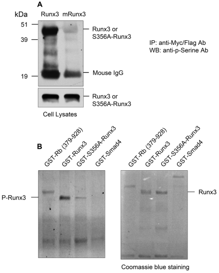
Fig. 3.
Cyclin D1 induces Runx3 phosphorylation. (A) COS cells were transfected with equal amounts of Myc-Runx3 and FLAG-S356A-Runx3 plasmids and treated with PS1 (10 μM, 4 hours incubation). Cell lysates were extracted 24 hours after transfection. Phosphorylation of Runx3 was detected by western blotting (WB) using the anti-phosphoserine antibody after WT and mutant Runx3 protein was immunoprecipitated (IP) using either the anti-Myc or anti-FLAG antibody. Strong phosphorylation was detected in WT Runx3-transfected cells. However, only trace amount of phosphorylated mutant Runx3 (P-Runx3) was detected. (B) In vitro phosphorylation assay. CDK4 was immunoprecipitated from 300 μg C2C12 cell lysate using 1.5 μg anti-CDK4 antibody and was used in the Runx3 kinase assay (1 μM substrate). GST-Rb (379-928) was used as a positive control and GST-Smad4 was used as a negative control. GST-Runx3 exhibits a strong phosphorylation, whereas GST-S356A-Runx3 exhibits a weak phosphorylation. Left: Runx3 phosphorylation by CDK4. Right: protein amounts, detected by Coomassie blue staining.

