Fig. 6.
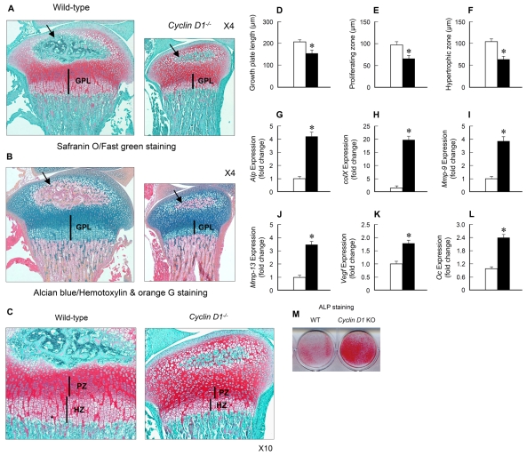
Chondrocyte proliferation and differentiation are altered in Ccdn1-knockout mice. (A-F) Histological analyses, including Safranin O/Fast green (A,C) and Alcian blue/Hemotoxylin and Orange G (B) staining, showed that growth plate length (GPL; D) and the lengths of the proliferating zone (PZ; C,E) and the hypertrophic zone (HZ; C,F) were reduced (n=6) and the formation of the secondary ossification center was delayed (black arrows in A and B) in Ccdn1-knockout mice compared to their WT littermates. (G-M) Primary chondrocytes were isolated from 3-day-old Ccdn1-/- mice and WT littermates and expression of chondrocyte marker genes and alkaline phosphatase (ALP) staining were examined. The results demonstrated that the expression of chondrocyte marker genes, such as Alp (G), collagen type X (ColX; H), Mmp9 (I), Mmp13 (J), Vegf (K) and osteocalcin (Oc; L), and ALP activity (M) were significantly increased in Ccdn1- knockout chondrocytes (n=3). *P<0.05, compared with the WT littermate controls; unpaired Student's t-test.

