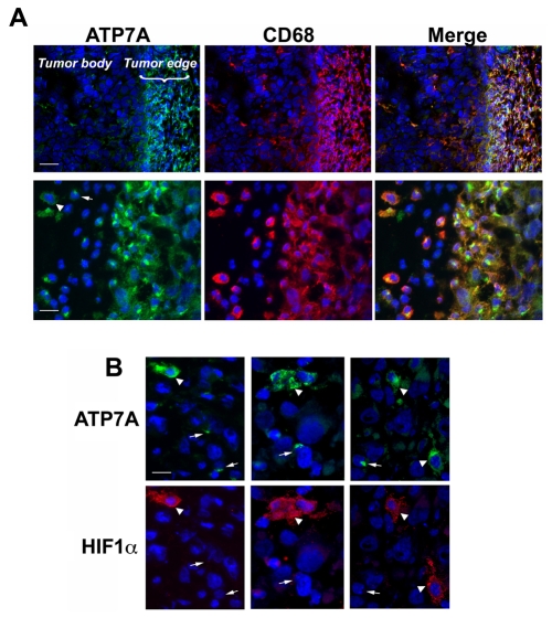
Fig. 3.
ATP7A trafficking in HIF1α-positive tumor-associated macrophages. (A) ATP7A is strongly expressed in tumor-associated macrophages. Human prostate cell PC-3 xenograft tumors from SCID mice were cryosectioned and probed with antibodies against ATP7A (green) or antibodies against the macrophage marker CD68 (red). Nuclei were stained using DAPI (blue). Upper panels show both the tumor mass and the tumor edge, with strong ATP7A expression in CD68-positive macrophages associated with the tumor edge Scale bar: 60 μm. The lower panels are higher magnifications of the tumor periphery and reveal extensive coexpression of ATP7A in CD68-positive macrophages, as indicated by yellow signal in the merged image. Scale bar: 30 μm. The arrow and arrowhead (lower left panel) indicate heterogeneous localization of the ATP7A in macrophages, in either a perinuclear or dispersed distribution. (B) The dispersed distribution of the ATP7A occurs in HIF1α-positive macrophages. Three different PC-3 tumor cryosections were immunostained for ATP7A (green) and HIF1α staining (red). ATP7A was restricted to the perinuclear region of macrophages negative for HIF1α expression (arrows), whereas a dispersed distribution of ATP7A occurred in HIF1α-positive macrophages (arrowheads) (original magnification, ×600). Scale bar: 10 μm.

