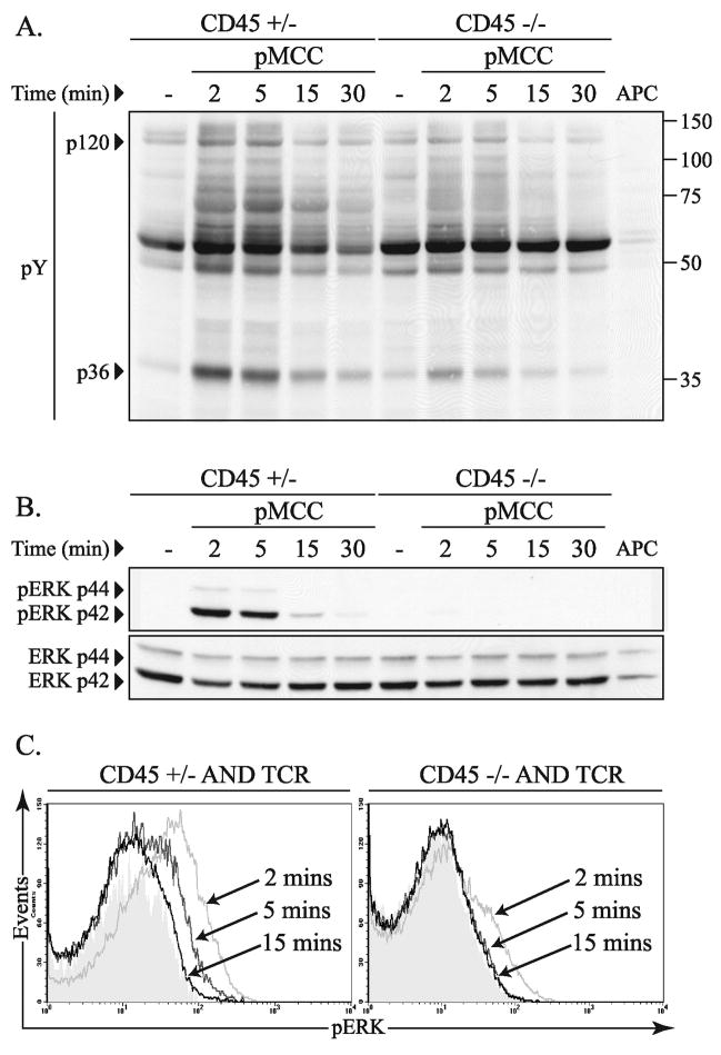
Figure 3.
Peptide dependent thymocyte activation in the absence of CD45. (A) Total thymocytes (2 × 10⁶) from CD45+/− or CD45−/− TCR transgenic mice were analyzed for induction of total tyrosine phosphorylation (pY) over time by western blot following stimulation with pMCC (50 μg/ml) pulsed T-depleted spleen cells as antigen presenting cells. (B) The pY blot shown in panel A was then stripped and probed for phosphorylated ERK (pERK) and repeated again for total ERK. The data shown are representative of three independent experiments. (C) Representative histograms of intracellular ERK phosphorylation in thymocytes from CD45+/− and CD45−/− mice following peptide stimulation for the indicated times. The cells were labeled with CD4 and CD8 in addition to antibodies to phosphorylated ERK. The data shown are gated on the double positive (CD4+, CD8+) cells and are representative of three independent experiments.

