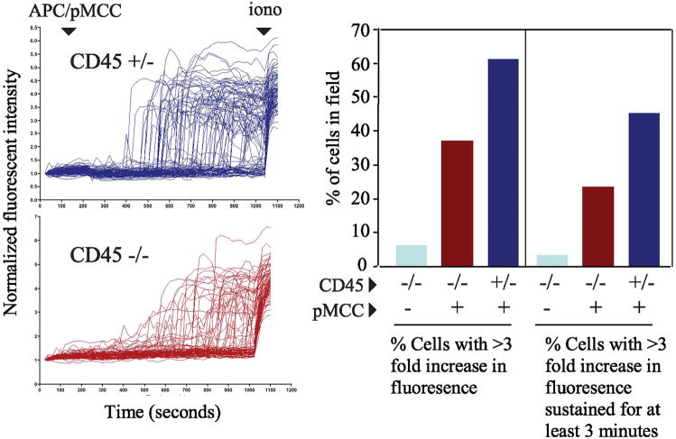
Figure 5.
Partial defect in sustained calcium mobilzation in individual CD45- double positive thymocytes following peptide stimulation. (A) Purified double positive thymocytes were loaded with 5μM fluo-4/AM ester and individual cell images were obtained every 20 seconds using the Biorad 1024 laser confocal microscope. After scanning was initiated 4 × 10⁶ APCs (T-depleted splenocytes) that were previously pulsed with 50 μg/ml pMCC peptide were added to the thymocytes. The initial average fluorescence of each cell was normalized to one and the results were expressed as changes in normalized fluorescence over time. Ionomycin was added at the end of scanning as a positive control to ensure that changes in calcium were detectable in the imaged cell population. (B) The percentage of responding cells was determined by dividing the number of cells in the field whose fluorescence increased 3-fold or was sustained 3-fold for more than 3 minutes by the total number of cells in the field. For CD45+ cells n=93, for CD45 deficient cells n=89. The data are representative of 2 independent experiments.

