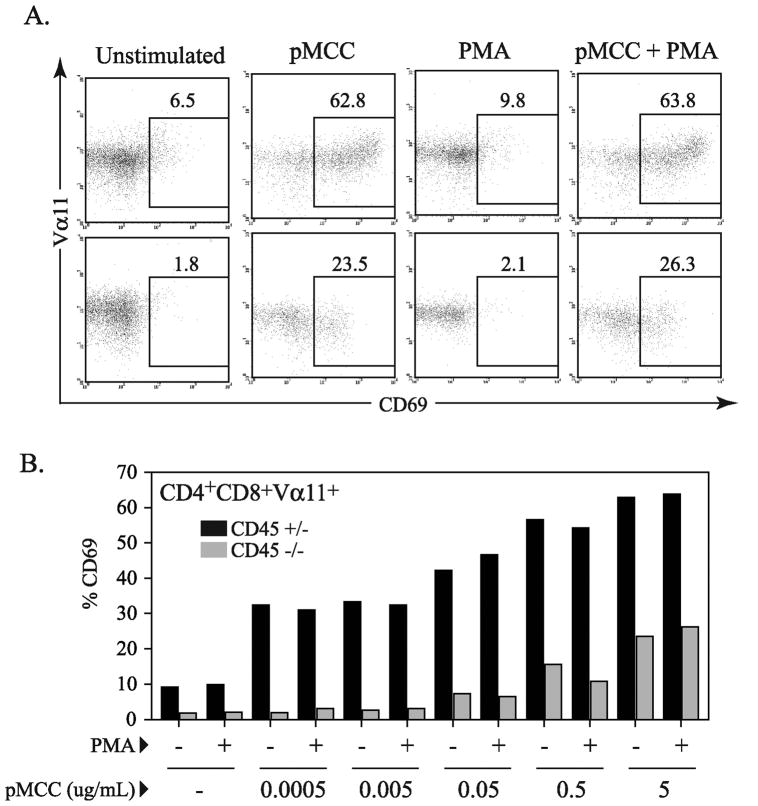
Figure 6.
Defect in CD69 induction in CD45 deficient thymocytes. (A) Representative dot plots displaying CD69 expression gated on CD4+CD8+ population after overnight stimulation with 5μg/mL pMCC and/or PMA at 0.1ng/mL. (B) Graph of %CD69 expression from flow cytometry data of thymocytes from CD45+/− or CD45−/− AND TCR transgenic mice after overnight stimulation in the presence of T cell depleted spleenocytes with varied concentrations of peptide (serial 10 fold dilutions with highest concentration at 5 μg/ml) and/or PMA at 0.1 ng/mL. All CD69 expression data presented is gated on CD4+CD8+Vα11+ population. This data is representative of three independent experiments.

