Figure 2.
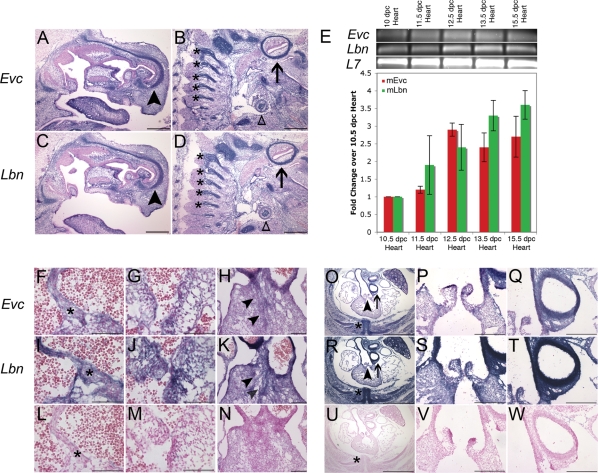
Expression of Evc and Lbn mRNA in the developing mouse embryo during AV septation. (A–D) Evc and Lbn mRNA expression in a sagittal section of a 15.5 dpc mouse whole embryo. Transcript for both genes is found in the cartilage primordium of the nasal bone (arrowhead), the vertebrae (astericks) and other cartilagenous structures like the primordium of the clavicle (open triangle) and the temporal bone (arrow). The scale bars in (A–D) represent 500 µM. (E) Quantitative RT–PCR confirms the presence of Evc and Lbn message in the developing murine heart from 10 dpc to 15.5 dpc. There is no statistically significant difference in the levels of Evc and Lbn mRNA transcript (P = NS). In situ hybridization in transverse sections of the heart at 13.5 dpc pinpoints co-expression at the tip of the primary atrial septum (asterisks in F and I), in the AV cushions (G and J) and in the connective tissue of the outflow tract (H and K). Sense controls are provided in (L–N). Transverse sections at 15.5 dpc reveal Evc and Lbn expression in structures affected in Evc syndrome including the heart (O–T) and the ribs (asterisks in O and R). Co-expression in the heart is strongest in outflow tract structures (O–T). The areas pointed out by the arrowhead in (O and R) are magnified in (P and S), while the arrows in (O and R) indicate the areas magnified in (Q and T). Sense controls are provided in (U–W). The scale bar represents 200 µM in all images from (F) to (W) except for (Q), (R) and (U), where they represent 1 mm.

