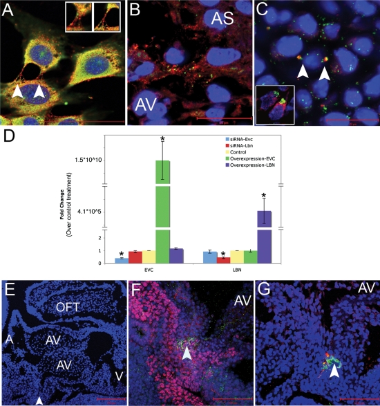
Figure 6.
EVC and LBN proteins are coexpressed in cell culture and in cells crucial for cardiac AV septation but there is no hierarchal transcriptional interregulation. EVC and LBN proteins are coexpressed in a similar pattern in NIH3T3 cells (A) and in vivo at the tip of the atrial septum (B) of a 12.5 dpc mouse. Arrows indicate overlapping expression of EVC and LBN in bone (C) and in the developing AV valve at 13.5 dpc (inset in C). Knockdown and overexpression of EvC-associated genes reveals a lack of hierarchial transcriptional interregulation (D). Manipulation of gene expression in cell culture revealed that changes in expression of EVC did not impact the levels of Lbn transcript and changes in the expression of LBN did not affect Evc transcription. The blue bars indicate successful knockdown of Evc after treatment with siRNA, but no change in Lbn gene expression. The red bars show Lbn could be knockeddown but the treatment did not affect Evc transcription. The yellow bar represents transfection with a control plasmid or a control siRNA. The green bar indicates successful overexpression of EVC with no affect on levels of Lbn transcription. The purple bar shows transfection of LBN does not alter the levels of Evc in the cell. Asterisks indicate statistical significance based on the Bonferroni adjusted P-value <0.0125. (E–G) are sagittal sections of a mouse heart at 10.5 dpc. AS, atrial septum; AV, atrioventricular cushions; A, atria; V, ventricles; OFT, outflow tract. The arrowhead in (E) indicates the area of zoom in (F) and (G). (F) shows overlap of ISL-1 (pink) and LBN (green) while G shows overlap of EVC (red) and LBN (green) in the DMP.

