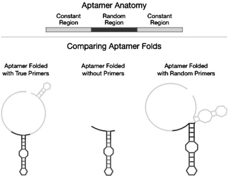
Fig. 1.

Cartoon schematic of the anatomy of an aptamer sequence. Top: Aptamer sequences consist of a random region (black rectangle) that is flanked by two constant regions (gray rectangles). The random region undergoes most evolution during in vitro selection, whereas the constant regions serve for amplifying the aptamers during the selection process. Bottom: Comparison of different folded strcutures used in the present study. This structure of the random region (black) is robust to the absence of constant regions or the presence of randomized constant regions (gray)
