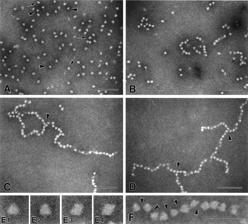
Figure 5.
Electron microscopy of crayfish CP samples. Negatively stained preparations of crayfish CP were analyzed by using electron microscopy. (A) CP monomers (CP monomers, HLS, and EDTA mixed); arrowheads indicate triangular projections; arrows indicate diamond-shaped projections. (B–D) CP monomers, HLS, and Ca2+ mixed, and the TGase-dependent crosslinking was stopped by addition of EDTA after 15 s (B), 45 s (C), and 60 s (D). Arrowheads in C and D indicate branchpoints. (A–D) Magnification ×172,000 (Bar = 100 nm.) (E) Selected triangular (E1 and E2) and diamond-shaped (E3 and E4) projections from the monomer preparation, magnification ×810,000. (F) Polymer of crosslinked CP molecules; arrowheads illustrate the deposition of stain between the individual CP molecules, indicating that the CP molecules interact at very localized points and that the interaction does not involve large surfaces of the individual CP molecules. Magnification ×515,000. (Bar = 50 nm.)

