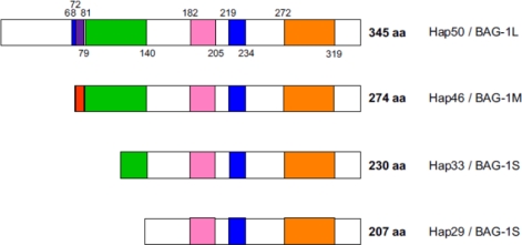
Figure 1.
Domain structure of polypeptides.Blue: potential nuclear localization signals; red: basic DNA-binding domain. The sequence in Hap46/BAG-1M is (1)Met-Lys-Lys-Lys-Thr-Arg-Arg-Arg-Ser-Thr(10). Due to overlap with a potential nuclear localization signal, this appears purple in Hap50/BAG-1L; green: acidic hexarepeat domain, consensus sequence: Thr-Arg-Ser-Glu-Glu-X; pink: ubiquitin-like domain; orange: hsp70/hsc70 binding domain or BAG-domain (shown here as originally defined [30,38]).

