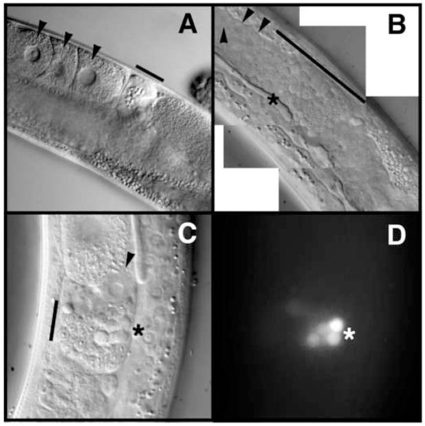
Figure 3.
Oogenesis and spermatogenesis defects in spr-5;amx-1 mutants. DIC microscopic imaging of the proximal gonad from wild-type (A) and severely sterile spr-5;amx-1 (B) adult worms. Black arrowheads point to oocytes (A) and defective looking oocytes (B and C). Black bars denote mature sperm in the spermatheca (A) as compared to early spermatogenic stages in the proximal gonad (B) and spermatheca (C). DIC imaging (C) and corresponding acridine orange staining (D) showing residual bodies (*).

