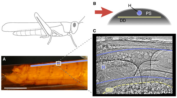
Figure 1.
Flow visualization in the heart of a grasshopper (Schistocerca americana) using synchrotron x-ray phase-contrast imaging. (A) Side view of the grasshopper abdomen showing the approximate location of the heart (blue) and the relative size of the imaging window (white rectangle, 1.3 × 0.9 mm). The abdomen is encapsulated in an x-ray transparent Kapton tube. Scale bar, 5 mm. (B), Cross-sectional schematic of the dorsal abdomen showing the relative sizes and locations of the heart (H), dorsal diaphragm (DD), and pericardial sinus (PS). The red arrow indicates the orientation of the x-ray beam. (C) X-ray video still of a region in the dorsal 3rd abdominal segment in lateral view. Round structures are air bubbles used to visualize patterns of heartbeat and hemolymph flow.

