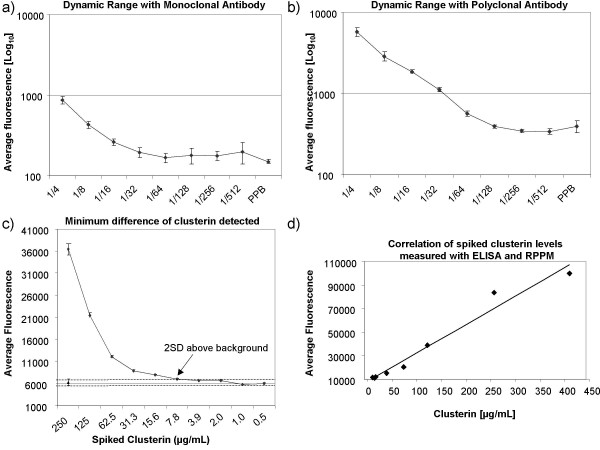
Figure 3.
Evaluation parameters of RPPMs. (a) Detection of clusterin in plasma samples using monoclonal and (b) polyclonal primary anti-clusterin antibodies. The log10 average signal intensity for clusterin was plotted against plasma dilution 1/4 to 1/512. (c) Minimum difference in clusterin concentration detected on RPPMs. Twenty different concentrations of recombinant clusterin were spiked in plasma and the average fluorescence intensity of three slides median normalized was plotted against the ten highest clusterin concentrations. The average fluorescence level (▲) with the value of 2 standard deviations (---) for the ten lowest endogenous clusterin concentrations are used as background clusterin level in this experiment. Arrow points to the concentration of spiked clusterin that yielded a signal at least 2SD above background plasma clusterin. (d) Correlation of clusterin levels measured with ELISA and RPPM (r = 0.989). Clusterin was measured in plasma samples spiked with increasing concentrations (0.9–500 μg/ml) of recombinant clusterin.

