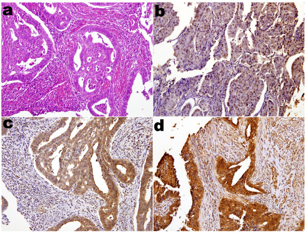
Figure 2.
Immunohistochemical analysis of p16INK4a staining in endometrial adenocarcinomas. (a) Photomicrograph revealed adenocarcinoma of endometrium, endometroid type, H&E stain. (b) Photomicrograph revealed tumor with more predominant p16INK4a staining at nuclei than that at cytoplasms. Diffusely moderately positive nucleic staining and no cytoplasmic staining were identified. (c) Photomicrograph revealed tumor with more predominant p16INK4a staining at cytoplasms than that at nuclei. Diffusely moderately positive cytoplasmic staining and focally weakly nucleic staining were identified. (d) Photomicrograph revealed tumor with dual prdominat p16INK4a staining at both cytoplasms and nuclei. Diffusely strongly positive cytoplasmic staining and nucleic staining were identified. All photomicrographs a, b, c, d were taken in median-powered, ×200.

