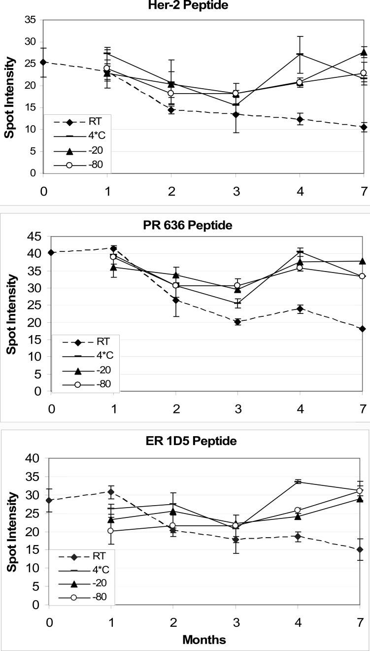
Figure 5.
Stability of peptide controls, for HER-2 (top panel), progesterone receptor (PR) 636 monoclonal antibody (middle panel), and estrogen receptor (ER) 1D5 monoclonal antibody (bottom panel). The dotted line represents the immunoreactivity of peptide controls stored at room temperature. The slight dip in immunoreactivity at the 3 month time interval is related to a slight inconsistency in the staining protocol (immunohistochemical detection) rather than an actual decrease in stability. Each time point is the mean ± SD of four replicate peptide controls.

