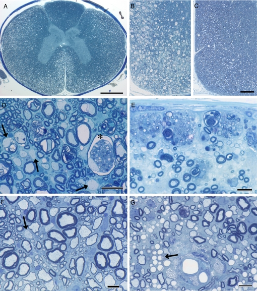
Fig. 1.
Demyelination and remyelination in the spinal cord. During acute disease, extensive changes are seen in the white matter of the spinal cord (A, B, D, and E). In the cervical cord, myelin vacuolation can be seen throughout the lateral and ventral columns (A and B) and on higher power (D). The deeper white matter and dorsal column is less affected (A). Vacuolation of myelin inevitably led to demyelination but with no loss of axons (D). In one myelinated axon, myelin debris is present next to an intact axon (*); other axons can be seen in adjacent fibers undergoing myelin vacuolization. There are numerous scattered remyelinated axons (thin myelin sheaths) and two demyelinated axons (D, arrows). In the dorsal column in a second cat, macrophages filled with myelin debris line the pia above numerous adjacent demyelinated axons. In marked contrast, a cat that had been fed a normal diet for 6 months showed almost complete myelin repair (C and F). Few vacuoles persisted, and the myelinated fiber density appeared normal (C). On higher power (F), it can be seen that many fibers of all diameters were remyelinated with only occasional demyelinated axons remaining (arrow). In 2 cats, although remyelination also was extensive in dorsal columns (G), numerous lipid-filled macrophages persisted adjacent to blood vessels, although many remyelinated axons were also present. There also appeared to be collections of capillaries adjacent to these macrophages (arrow). Toluidine blue. (Scale bars: A, 1.0 mm; B and C, 200 μm; D, 20 μm; E–G, 10 μm.)

