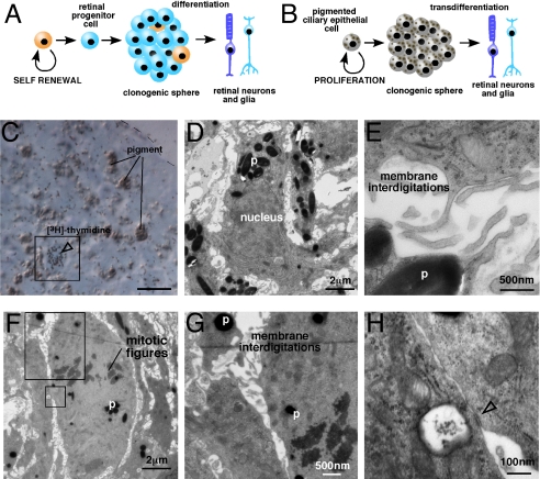
Fig. 3.
Pigmented CE cells are proliferating in CE-derived spheres. (A and B) Comparision of the retinal stem cell model and the transdifferentiation model. (C) S-phase cells were labeled by incubating Day 3 spheres with [3H]-tymidine for 1 h. Immediately after labeling, the spheres were fixed and a 1-μm thick section was collected from the center of the sphere. This section was stained with toluidine blue and overlaid with autoradiographic emulsion to detect the cells in S-phase (box, open arrowhead). (D and E) Immediately after the 1-μm section, 10 serial 50-nm sections were collected for transmission electron microscopy. These sections were imaged and aligned to the [3H]-tymidine–labeled section to characterize the morphology of the cells in S-phase. The cell shown in (C) is also shown in (D and E) by aligning the sections. (F–H) Cells in M-phase were identified by the presence of condensed chromosomes (mitotic figures). These cells also contained pigment, membrane interdigitations, and epithelial junctions (open arrowhead). Abbreviations: p, pigment. (Scale bar in C, 10 μm.)

