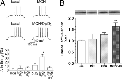
Fig. 2.
Interaction of the MCH1R with D1R and D2R in the NAcSh. (A) Changes in NAcSh spike firing induced by MCH (2.5 μM) alone, D1 agonist (SKF 81297 3 μM) plus MCH, D2 agonist (Quinpirole 3 μM) plus MCH, D1+D2 agonists, D1+D2 agonists plus MCH, and D1+D2 agonists plus MCH and TPI 1361-17 (2 μM) (*, P < 0.05 for MCH/D1/D2 vs. each other condition, F (5, 25) = 6.66; *, P < 0.001, for 1-way ANOVA, followed by Bonferroni multiple comparison test; n = 4–6). Resting potential for MCH example was −82.4 and −83.2 mV before and after MCH, and resting potential for MCH/D1/D2 example was −80.2 and −79.6 mV before and after agonists. (B) Phosphorylation of DARPP-32 (Thr 34) in NAcSh slices after MCH (2.5 μM) alone, D1+D2 agonists (SKF 81297 3 μM and Quinpirole 3 μM), and D1+D2 agonists plus MCH (**, P < 0.01 for MCH/D1/D2 vs. control, ANOVA followed by Bonferroni multiple comparison test; n = 6–10). Immunoblots for detection of phospho-Thr-34 DARPP-32 are shown at the top. The levels of phospho-Thr-34 DARPP-32 were normalized to values obtained from control slices (a.u., arbitraty unit).

