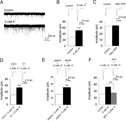
Fig. 4.
Activity-induced trafficking of AMPAR requires activation of PKCγ. (A) Representative recordings of mEPSC before and after application of 5 mM K+. (B) Application of 5 mM K+ for 10 min significantly increased mEPSC amplitude (n = 5). (C) Induction of postsynaptic depolarization of the Mauthner cell via a DPP had no effect on the mEPSC amplitude (n = 5). (D) Intracellular application of γV5-3 (10 nM) prevented the potentiation in amplitude after the 5 mM K+ bath (n = 5), whereas the control peptide (C1; 10 nM) had no effect (n = 5). (E) Inclusion of pep2m (200 μM, n = 5) in the recording solution completely prevented the 5 mM K+ induced increase in mEPSC amplitude, whereas the control peptide, pep4c (200 μM, n = 5) had no effect. (F) Application of APV (50 μM, n = 7) completely blocked the 5 mM K+ induced increase in mEPSC amplitude. ***, significantly different, P < 0.001.

