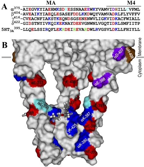
Fig. 9.
An azietomidate binding pocket in the nAChR cytoplasmic domain. A, an alignment of the sequences of the T. californica nAChR subunit and human 5-HT3A receptor MA/M4 helices, with the positions in the nAChR α subunit photolabeled by [3H]azietomidate, [3H]azicholesterol (Hamouda et al., 2006), and [3H]azioctanol (Pratt et al., 2000) colored cyan, purple, and brown, respectively. Other acidic and basic amino acids are colored red and blue, respectively. The positions in the 5-HT3A receptor identified as conductance determinants are green (Hales et al., 2006). B, a Connolly surface representation of the basket formed by the MA helices, viewed from the side with the β and δ subunits removed to visualize the interior and with the amino acids color-coded as in (A). An azietomidate molecule in stick format is included docked in its lowest energy orientation (see Materials and Methods).

