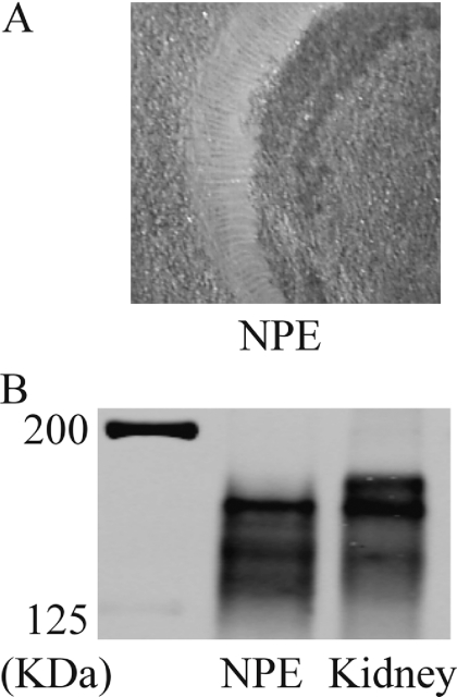
Fig. 2.
A, image showing the freshly dissected porcine NPE. The tissue is opaque in color, whereas the background is tan. Pigmented cells and vascular tissue are not discernable. B, Western blot showing expression of MRP2 in the native porcine NPE and porcine renal cortex. Twenty-five micrograms of NPE lysate and renal cortex (kidney) crude homogenate was used for electrophoresis.

