Figure 3.
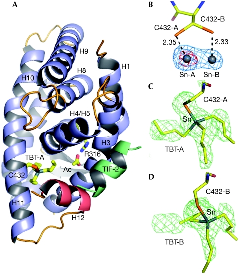
Structure of the RXR-α ligand-binding domain in complex with TBT and TIF2. (A) Overall structure of the complex in cartoon representation. The TBT is shown bound to Cys432 through a covalent interaction, and an acetate molecule (Ac) involved in a salt bridge with Arg316 are depicted. This acetate, not essential for RXR activity, derives from the crystallization condition. (B) Anomalous difference electron density map contoured at 15.0σ (red) and 5.0σ (blue) showing two tin sites facing the two alternative positions of Cys432 (A,B). (C,D) The two positions of TBT (TBT-A and TBT-B) bound to Cys432-A and Cys432-B are shown in the FO–FC omit map contoured at 3.0 σ. LBD, ligand-binding domain; RXR-α, retinoid X receptor-α; TBT, tributyltin; TIF2, transcriptional intermediary factor 2.

