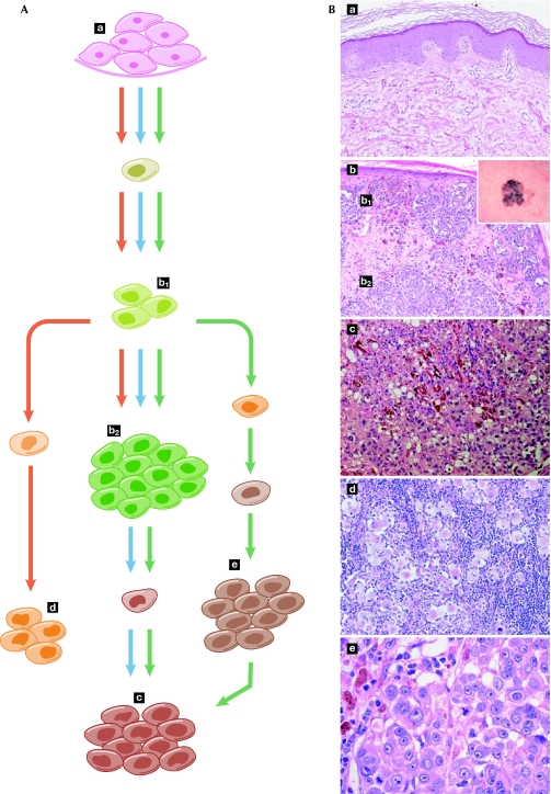
Figure 3.
Models of cancer evolution. (A) The ‘clonal selection model' (blue arrows) is the prevailing view to explain the successive steps of mutation and selection from normal tissue to primary tumour and metastasis. However, metastasis-generating cells can emerge relatively early in the tumorigenic process and ‘seed' distant tissues, thereby evolving in parallel with the primary tumour and delineating the ‘parallel evolution' model (red arrows). Finally, these two models can occur simultaneously and metastatic deposits can act as sites from which additional metastases can be generated, therefore leading to an integrated model of cancer evolution (green arrows). (B) Microphotographs provide a histological snapshot of normal skin tissue (a), primary tumour (superficial b1 and deep b2, macroscopic appearance inset in b), subcutaneous metastasis (c), metastasis in the lymph node (d) and metastasis in the lung (e), and are shown in correspondence with the cancer-evolution models. This melanoma—which originates from the transformation of pigmented skin cells—provides a visual example of the modelling paradigms, illustrating the gap between ideal models and actual observations.

